Diagram Of The Muscles In The Forearm : If You Pop Stop Recognizing A Bicep Tendon Tear Angie S List - The muscles in the flexor compartment are mainly innervated by the musculocutaneous nerve , while the extensors are innervated by the radial nerve.
Diagram Of The Muscles In The Forearm : If You Pop Stop Recognizing A Bicep Tendon Tear Angie S List - The muscles in the flexor compartment are mainly innervated by the musculocutaneous nerve , while the extensors are innervated by the radial nerve.. Diagram of the forearm flexors. From the arm muscle diagram above, the muscles of the arm that can be seen easily on the surface include biceps, triceps, brachioradialis, extensor carpi radialis longus, and deltoid. The muscles of the arm are responsible for the movement of your elbow and, by extension, your forearm. This muscle has two heads. They control movements of the wrist, hand, fingers and thumb.
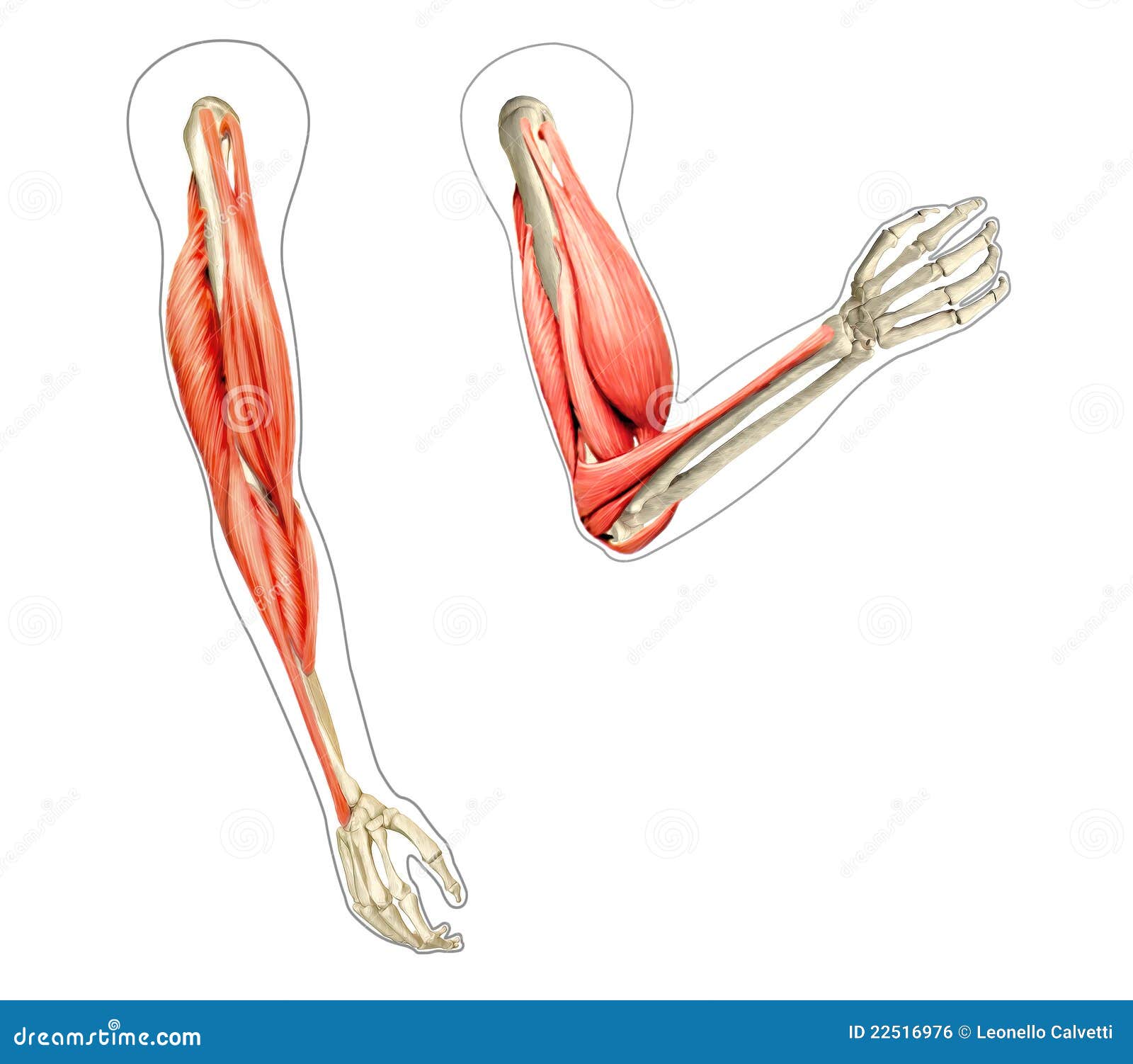
Rotates the forearm so the palm is facing the ceiling). Human muscle system, the muscles of the human body that work the skeletal system, that are under voluntary control, and that are concerned with movement, posture, and balance. The photo on the left shows muscles that are deep to the ones on the right. Five muscles originate on either the humerus or the scapula and insert onto the bones of the forearm to flex and extend the elbow: The triceps brachii muscle is the prime extensor of the forearm at the elbow joint, with assistance from the anconeus muscle, but is also capable of weak arm extension and adduction.
Deep fascia of the forearm).—the antibrachial fascia continuous above with the brachial fascia, is a dense, membranous investment, which forms a general sheath for the muscles in this region;
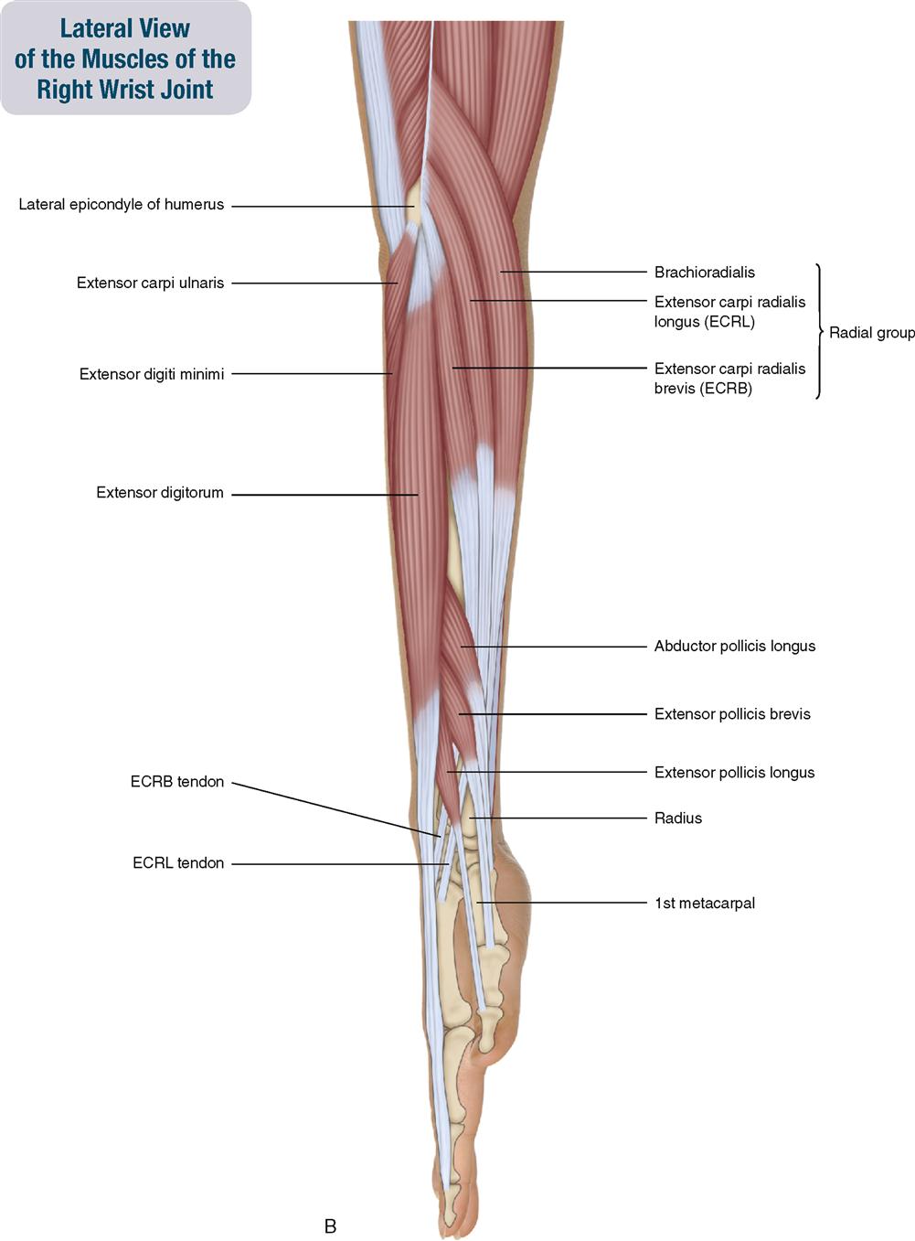
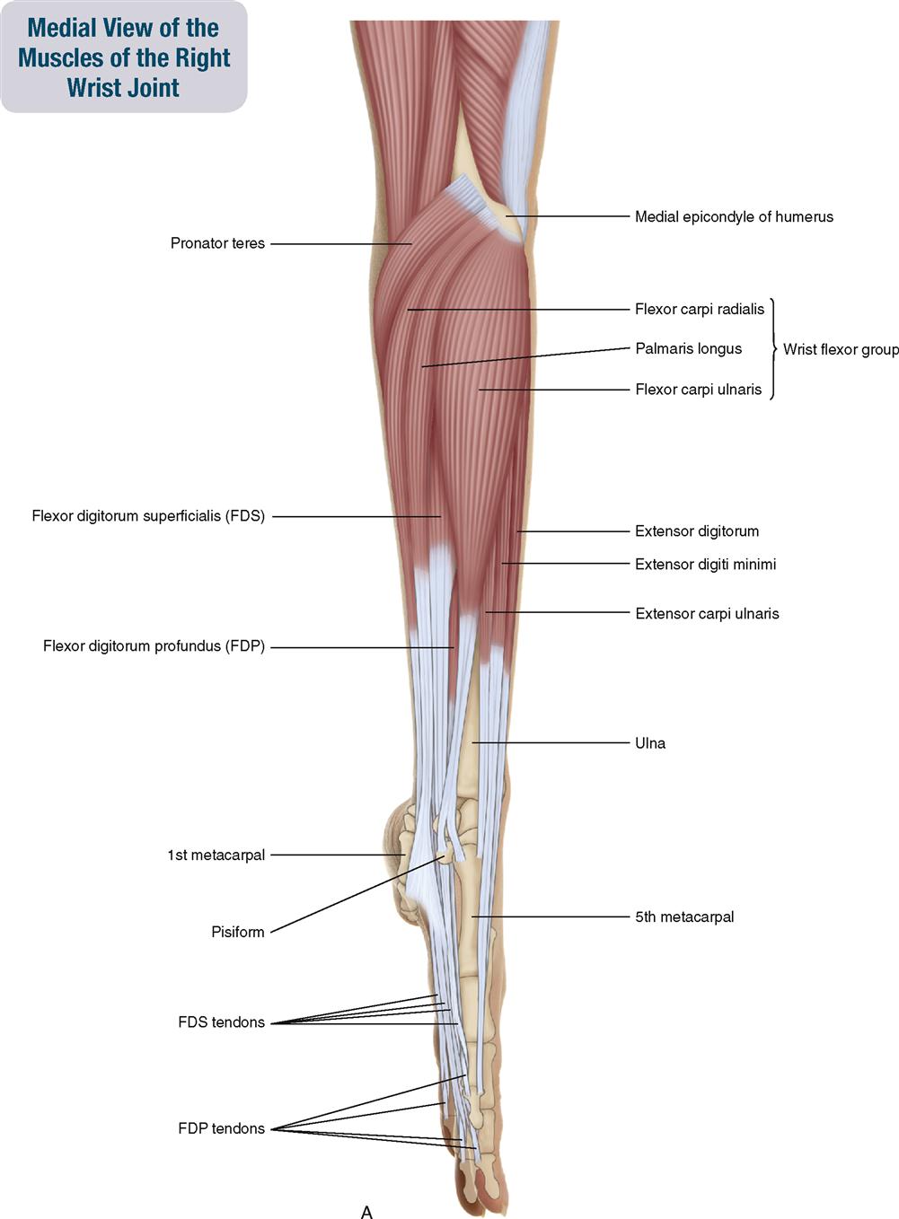
Learn vocabulary, terms, and more with flashcards, games, and other study tools. Flexes elbow and moves forearm. The short head originates on the … This muscle has two heads. Human anatomy lever 12 photos of the human anatomy lever human anatomy levers, human muscles, human anatomy levers. Deep fascia of the forearm).—the antibrachial fascia continuous above with the brachial fascia, is a dense, membranous investment, which forms a general sheath for the muscles in this region; As seen in this forearm muscles diagram, the flexor muscles reside in the anterior compartment of the forearm, and are … The arm is one of the body's most complex and frequently used structures. Rotates the forearm so the palm is facing the ceiling). The tendons of the hand extensor muscles pass under the extensor retinaculum and attach to the. The superficial extensors of the forearm are the brachioradialis, extensor carpi radialis longus, anconeus, extensor carpi radialis brevis, extensor carpi ulnaris, extensor digitorum and extensor digiti minimi. Yoga anatomy anatomy study anatomy reference anatomy bones anatomy drawing hand therapy massage therapy physical therapy occupational therapy. Biceps are large muscle of the upper arm is formally known as the biceps brachii muscle, and rests on top of the humerus bone.
(the lower arm is the forearm or antebrachium.) there are three muscles on the upper arm that are parallel to the long axis of the humerus, the biceps brachii, the brachialis, and the triceps brachii. The forearm muscles that control the movement of the hands are known as extrinsic hand muscles. Grade iii strain of forearm muscle: It is the most superficial muscle of the radial side of the forearm, forming the lateral wall of the cubital fossa. Most of these originate from the lateral epicondyle.
By darkreign plays quiz not verified by sporcle.
These types of strain are moderate in nature in that there is tearing of fibers in the muscle or tendons at its attachment to the bone. When the biceps contracts, it pulls the forearm up and rotates it outward. Broadly considered, human muscle—like the muscles of all vertebrates—is often divided into striated muscle, smooth muscle, and cardiac muscle. The muscles that extend the hand at the wrist are located on the posterior portion of the forearm. We are pleased to provide you with the picture named right arm muscle and tendon anatomy.we hope this picture right arm muscle and tendon anatomy can help you study and research. The tendon that attaches the biceps muscle to the forearm bones (radius and ulna) is called the distal biceps tendon. Muscles of the ant/ventral forearm: These types of strains are quite severe and involve complete rupture of the muscle fibers and tendons. By darkreign plays quiz not verified by sporcle. The short head originates on the … This muscle flexes the elbow and shoulder as well as supinates the forearm (i.e. These muscles originate outside the hand and insert on structures within it. The superficial extensors of the forearm are the brachioradialis, extensor carpi radialis longus, anconeus, extensor carpi radialis brevis, extensor carpi ulnaris, extensor digitorum and extensor digiti minimi.
The tendons of the hand extensor muscles pass under the extensor retinaculum and attach to the. The tendon that attaches the biceps muscle to the forearm bones (radius and ulna) is called the distal biceps tendon. These muscles originate outside the hand and insert on structures within it. Grade ii strain of forearm muscle: Learn with flashcards, games, and more — for free.
The muscles that extend the hand at the wrist are located on the posterior portion of the forearm.
Start studying muscles of the arm. Biceps are large muscle of the upper arm is formally known as the biceps brachii muscle, and rests on top of the humerus bone. The biceps brachii is on the anterior side of the humerus and is the prime mover (agonist) responsible for flexing the forearm. Science quiz / arm and forearm muscles random science or anatomy quiz can you pick the arm and forearm muscles? From the arm muscle diagram above, the muscles of the arm that can be seen easily on the surface include biceps, triceps, brachioradialis, extensor carpi radialis longus, and deltoid. This muscle has two heads. When the biceps contracts, it pulls the forearm up and rotates it outward. The superficial extensors of the forearm are the brachioradialis, extensor carpi radialis longus, anconeus, extensor carpi radialis brevis, extensor carpi ulnaris, extensor digitorum and extensor digiti minimi. Diagram of the forearm flexors. It is the most superficial muscle of the radial side of the forearm, forming the lateral wall of the cubital fossa. Rotates the forearm so the palm is facing the ceiling). The long head originates just above the shoulder socket on the scapula and blends with the short head onto the radius bone of the forearm. We'll go over all the muscles in your upper arm and forearm as well as explain.


Komentar
Posting Komentar